The Importance of Radiology in the Diagnosis and Treatment of Heart Diseases
A faculty member of Iran University of Medical Sciences explained the importance of medical imaging in the diagnosis and treatment of heart diseases.


A faculty member of Iran University of Medical Sciences explained the importance of medical imaging in the diagnosis and treatment of heart diseases.
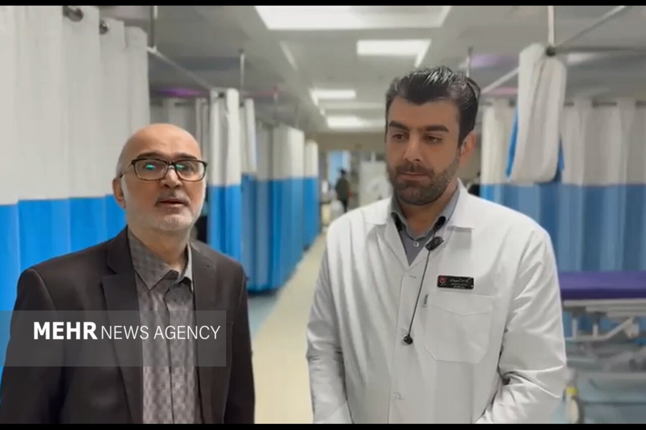
According to Mehr News Agency, Hamidreza Pourali-Akbar, a faculty member of Iran University of Medical Sciences, said on the eve of the 41st Iranian Congress of Radiology that the main theme of this year's congress is cardiac imaging: “At this congress, compared with previous editions, special attention has been paid to this field and we will have a particular focus on cardiac topics; topics which, because they were not the main theme before, we used to address less.”
The radiology specialist, noting that the congress has 12 main tracks, added: “In the track related to the congress's main theme, namely the heart, we have numerous sessions and lectures in the fields of coronary CT angiography, CT angiography of the great vessels, and structural heart diseases; that is, the procedures carried out in our country toward non-surgical interventions. Our goal is to enhance the skills of trainees and participants.”
The secretary of the 41st Iranian Congress of Radiology also announced a session on cardiac MRI, saying: “In this session we will address diseases of the heart muscle and the findings that can be pointed to in cardiac MRI, in the form of refresher training and the transfer of the valuable experiences and points that our colleagues have.”
Online session with one of the world's best CT angiography instructors
Regarding the congress's educational programs, Pourali-Akbar said: “To raise our colleagues' scientific ability, we have several dedicated sessions examining imaging of cardiovascular diseases. One of these special sessions is held online with one of the world's best instructors of coronary CT angiography, so that they teach the newest and latest findings to our colleagues.”
The role of imaging in the prevention, diagnosis and treatment of heart diseases
On the place of cardiac imaging, he stated: “Advances in methods such as CT angiography and cardiac MRI have made it possible to examine the coronary arteries, heart muscle, valves and great vessels more precisely, and help physicians choose the appropriate treatment method.”
Pourali-Akbar said: “Early identification of disease and control of risk factors are among the most important strategies for reducing cardiovascular complications. Coronary CT angiography, by non-invasively examining the vessels feeding the heart, can help identify atherosclerotic plaques and vascular stenoses. Identifying these changes, alongside clinical assessment, can pave the way for better control of factors such as diabetes, high blood pressure and blood lipid disorders.”
He acknowledged: “Performing CT angiography is not necessary for everyone. The choice of this method or other methods of examining the coronary arteries must be made based on symptoms, medical history, the level of disease risk and the physician's opinion. Consulting experienced specialists in heart diseases and cardiac imaging helps in choosing the appropriate method and avoiding unnecessary tests. Medical imaging, although useful, must be purposeful and proportionate to each patient's needs.”
According to Pourali-Akbar, the application of cardiac imaging is not limited to the coronary arteries. CT angiography is important in evaluating aortic diseases, including dilation and aneurysm, and also in examining some pulmonary artery diseases.
He added: “This method is also used in examining congenital diseases of the heart and the great vessels. Some anomalies may exist from childhood and be diagnosed at older ages. Cardiac CT, by precisely displaying the structure of the heart and the origin and course of the coronary arteries, the aorta and the pulmonary arteries, can help diagnose complex anomalies and plan surgery or therapeutic interventions. The choice of CT or MRI in these patients depends on the type of anomaly, the patient's age and the clinical question.”
The radiology specialist said: “Cardiac MRI is also one of the important methods for examining the function and tissue of the heart muscle. The information obtained from MRI helps in more precise diagnosis and in choosing the appropriate treatment strategy. Another important advance in medicine is replacing the aortic valve via catheter, without the need for open surgery, in selected patients. In this method, called TAVI or TAVR, the new valve is guided to the heart through the vessels. Performing CT angiography before this procedure is very important for precisely measuring the valve and the aortic root, examining the openings of the coronary arteries and evaluating the catheter entry route, and it helps in choosing the type and size of the valve and reducing the risk of complications.”
He continued: “When a new technique enters the country, imaging technologies must also advance so that they can assist cardiologists and cardiac surgeons, including surgeons of congenital heart diseases, and help them reach the desired result. Therefore, this field moves in step with the advances of other branches of cardiology, and we hope this trend continues ever better.”
Equipment, despite limitations, still on the path of being updated
Pourali-Akbar said: “In order to provide appropriate services in cardiovascular imaging to the patient community and to the country's medical-science trainees, we have two important principles before us: first, up-to-date medical equipment, and second, the knowledge of how to use these tools correctly.”
Regarding equipment, he also stated: “The country's current position in equipment is acceptable, although there is a need for very serious effort to upgrade. With the help of the Radiology Association and the Ministry of Health, over the past two or three years a large number of the needed devices, including CT scan machines capable of CT angiography of the heart and coronary vessels and MRI machines capable of performing cardiac MRI, have entered the country.”
According to the secretary of the 41st Iranian Congress of Radiology, despite sanctions and currency fluctuations, supplying the newest technologies in the recent period has been accompanied by challenges, but efforts to procure and update equipment continue.
He added: “I hope that by removing some obstacles we can have maximum access to the most up-to-date diagnostic equipment and technologies in the country.”
Fellowship in cardiovascular imaging
Pourali-Akbar, referring to the 8 years since the launch of the cardiovascular imaging fellowship, said: “For a year and a half, the fellows are trained focusing only on the specialized details of the science and knowledge of this field.”
He added: “We have tried to use more of the young faculty members of the universities so that they can be a source of good in the big universities and other places in the country, from Shiraz, Tabriz, Isfahan and Mashhad to even some smaller universities. This work helps spread a knowledge that is necessary, completely up-to-date and in need of special technologies, and increases the range of radiologists' awareness in this field. In my opinion this is a very great event that is gradually spreading across the country and will greatly help our compatriots.”
Fetal imaging in the spotlight of the radiology congress
Behnaz Moradi, scientific secretary of the 41st Annual Iranian Congress of Radiology, also, emphasizing the growing importance of prenatal imaging, spoke of the role of advanced imaging methods, artificial intelligence and knowledge exchange with international experts in improving the diagnosis of fetal anomalies.
Referring to the importance of imaging during pregnancy, she stated: “Fetal imaging is one of the specialized and advancing fields of radiology that plays an important role in the prenatal diagnosis of structural anomalies, especially brain and heart disorders. The importance of this field is that decisions based on the results of fetal imaging are not limited to a single individual, but directly affect the health of the fetus and the mother, and in fact the future of a family. Therefore, precise and timely diagnosis, together with specialized consultation and informed decision-making, is of special importance and can help in appropriate planning for continuing the pregnancy, specialized prenatal care and providing optimal conditions for treating the newborn after birth.”
Moradi added: “At this year's congress, alongside joint interdisciplinary lectures and panels, numerous specialized workshops for fetal imaging have also been planned.”
Online fetal neuroimaging conference with international professors
The scientific secretary of the congress considered the holding of international scientific programs an important part of this event and said: “One of the congress's special programs is the online fetal neuroimaging conference, held with the presence of prominent international professors. This program provides an opportunity for Iranian specialists to become acquainted with the latest views, clinical experiences and scientific advances in the field of fetal brain imaging.”
Moradi added: “In this program, specialized topics related to evaluating the fetal brain, new imaging methods and diagnostic challenges are addressed. The presence of well-known international professors, in addition to raising the scientific level of the program, paves the way for exchange of experience, familiarity with the world's current approaches and strengthening scientific ties between Iranian specialists and the international radiology community.”
Joint interdisciplinary panels
One of the important approaches in the scientific planning of this edition of the congress was special attention to joint interdisciplinary panels. These panels, with the participation of radiology specialists and other related fields, including perinatology and pediatric specialists, make it possible to examine diagnostic and therapeutic challenges from different viewpoints.
Fetal heart echocardiography
Emphasizing the importance of examining the fetal heart, Moradi said: “Congenital heart diseases are among the most important congenital anomalies, and their prenatal diagnosis can play a decisive role in planning pregnancy care and managing the newborn after birth.”
She continued: “Specialized fetal heart echocardiography, performed by pediatric cardiology subspecialists and radiology specialists with knowledge of fetal heart echo, makes it possible to examine cardiac structures, valve function, blood flow and heart rhythm more precisely. Identifying some heart diseases before birth provides the opportunity to plan delivery in equipped centers and the presence of specialized pediatric and neonatal cardiology teams. Therefore, improving imaging quality and increasing diagnostic accuracy in this field is of special importance in mother and fetus care.”
Artificial intelligence and new technologies in fetal imaging
On the role of artificial intelligence in the future of medical imaging, Moradi stated: “Artificial intelligence is creating a transformation in many fields of radiology, and fetal imaging also benefits from these advances. Deep-learning-based technologies can play a role in improving image quality, identifying and separating anatomical structures, standardizing measurements and helping more precise analysis of imaging data.” According to her, in various congress sessions related to fetal imaging, the latest achievements and applications of artificial intelligence are addressed.
Referring to the challenge of fetal movement, she noted: “Fetal movement is one of the important limitations of fetal MRI, which sometimes reduces image clarity. New image-reconstruction methods, reducing motion artifacts and increasing image resolution using artificial intelligence algorithms, have considerable capacity to upgrade image quality. These advances can facilitate the evaluation of delicate fetal brain structures and some complex anomalies.”
Moradi emphasized: “The ultimate goal of using these technologies is not to replace the physician, but to provide more precise tools to increase confidence and speed of diagnosis and to help clinical decision-making.”
Specialized ultrasound and fetal MRI
The scientific secretary of the congress said: “In recent years, advances in specialized ultrasound, three-dimensional ultrasound and fetal MRI have made it possible to examine anatomical structures more precisely and evaluate some complex anomalies. Fetal MRI, especially in examining the central nervous system, in cases where ultrasound alone does not answer the diagnostic questions, can provide valuable supplementary information.”
The congress: a platform for linking knowledge, technology and clinical experience
In conclusion, Moradi, referring to the importance of holding scientific congresses, stated: “Medical radiology is transforming at great speed, and keeping pace with these advances requires continuous education, research, interdisciplinary cooperation and knowledge exchange with the world's reputable scientific centers.”
She added: “We hope the 41st Annual Iranian Congress of Radiology, by providing a platform for presenting the latest scientific achievements, examining new technologies and dialogue among domestic and international specialists, will be an effective step in raising the quality of the country's medical imaging services.”
The 41st Annual Iranian Congress of Radiology will be held from November 3 to 6, 2026, at the Iran Mall International Conference Center. This scientific event, with the wide presence of radiologists and other related specialists, provides an opportunity to exchange experiences, examine the latest achievements of medical imaging and become acquainted with the newest scientific advances in this field. Cardiac imaging and pregnancy imaging are the two main themes of this year's congress.

Source: Mehr News Agency
Related stories
The Latest Details on the Service-Delivery Status of Nabi Akram (PBUH) Hospital in Zahedan

Early Detection; An Effective Factor in Treating Stomach Cancer
