Updating Radiology Equipment Despite the Imposition of Sanctions
A modern news platform — accurate, fast and unbiased reporting
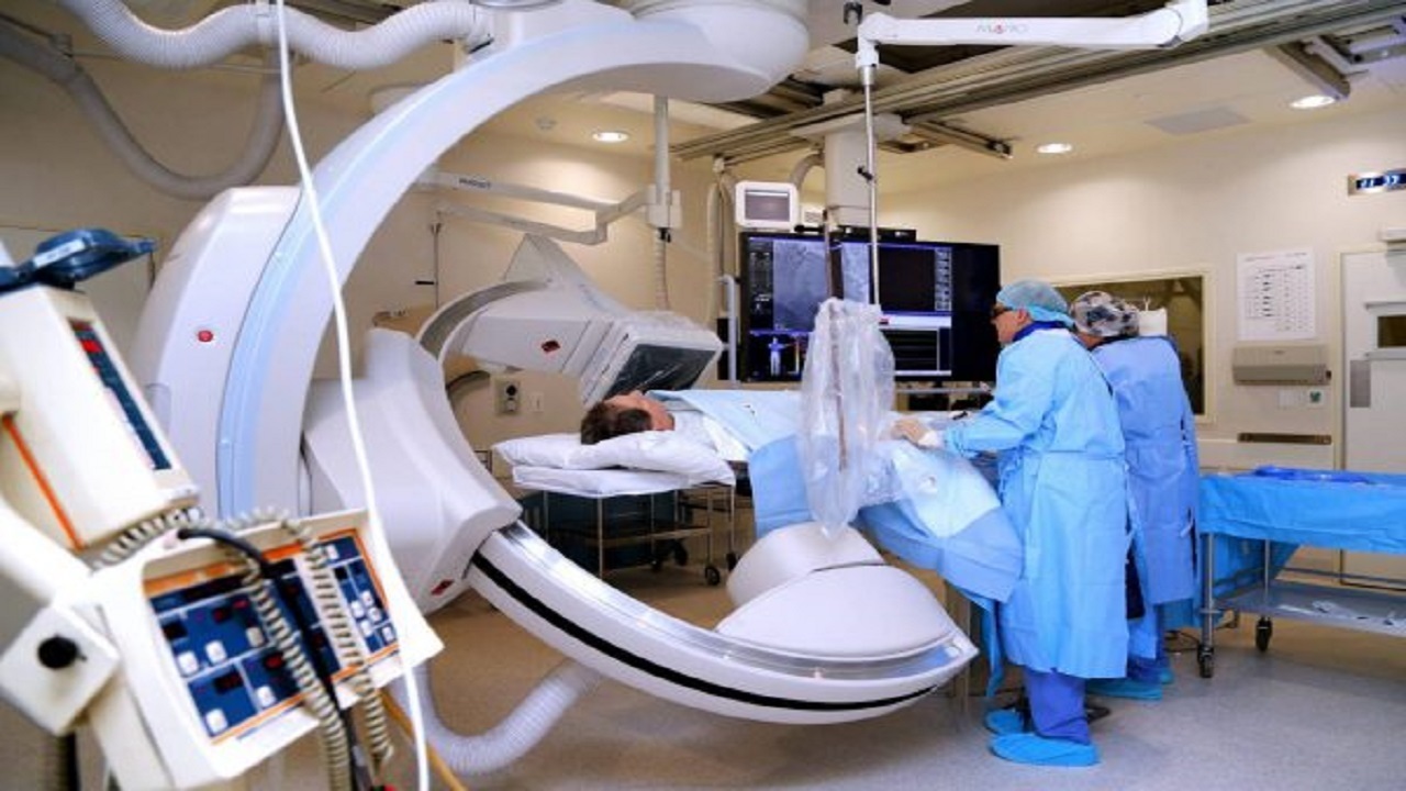
A faculty member of Iran University of Medical Sciences said: The country's radiology equipment is being updated despite the imposition of the enemies' sanctions.
Young Journalists Club - Hamidreza Pourali-Akbar, the congress secretary, a radiology specialist and faculty member of Iran University of Medical Sciences, pointing out that cardiac imaging is the main axis of this year's radiology congress, said: In this congress, compared with past congresses, special attention has been paid to this field, and we will have a particular focus on cardiac topics; topics which before, since they were not the main theme, we addressed less.
He, pointing out that the annual radiology congress has 12 main axes, added: In the axis that is related to the congress's main theme, namely the heart, we have numerous sessions and lectures in the field of coronary CT angiography, CT angiography of the great vessels and structural heart diseases (Structural Heart Disease); that is, the measures that are carried out in our country toward interventions without surgery. Our goal is to increase the capability of the learners and participants.
The congress secretary also announced the holding of a session about cardiac MRI and said: In this session we address the diseases of the heart muscle and the findings that can be pointed to in cardiac MRI; in the form of retraining and the transfer of the valuable experiences and points that exist with colleagues.
Dr. Pourali-Akbar, about the congress's educational programs, said: To increase the scientific capability of colleagues, we have several dedicated sessions for examining the imaging of cardiovascular diseases. One of these special sessions is online and held with one of the best world instructors of coronary CT angiography, so that they teach the new and novel findings to colleagues.
A faculty member of Iran University of Medical Sciences said: The country's radiology equipment is being updated despite the imposition of the enemies' sanctions.
The Role of Imaging in the Prevention, Diagnosis and Treatment of Heart Diseases
Dr. Pourali-Akbar, about the place of cardiac imaging, stated: The advancement of methods such as CT angiography and cardiac MRI has provided the possibility of a more precise examination of the coronary vessels, the heart muscle, the valves and the great vessels, and helps physicians in choosing the appropriate treatment method. Early identification of disease and control of risk factors are among the most important strategies for reducing cardiovascular complications. Coronary CT angiography can, by the non-invasive examination of the vessels feeding the heart, help identify atherosclerotic plaques and vascular stenoses. The identification of these changes, alongside clinical assessment, can provide the ground for better control of factors such as diabetes, high blood pressure and blood lipid disorders.
He acknowledged: Performing CT angiography is not necessary for all individuals. The choice of this method or other methods of examining the coronary vessels must be made based on symptoms, medical history, the level of disease risk and the physician's opinion. Consultation with experienced specialists in heart diseases and cardiac imaging helps in choosing the appropriate method and avoiding unnecessary tests. Medical imaging, although useful, must be targeted and proportionate to the need of each patient.
According to Dr. Pourali Akbar, the application of cardiac imaging is not limited to the coronary vessels. CT angiography has importance in evaluating aortic diseases, including dilation and aneurysm, and also in examining some pulmonary artery diseases.
He said: This method also has application in examining congenital diseases of the heart and great vessels. Some anomalies may exist from childhood and be diagnosed at older ages. Cardiac CT can, by precisely displaying the structure of the heart and the origin and course of the coronary vessels, the aorta and the pulmonary arteries, help in diagnosing complex anomalies and in planning for surgery or therapeutic interventions. The choice of CT or MRI in these patients depends on the type of anomaly, the patient's age and the clinical question.
He added: Cardiac MRI is also one of the important methods for examining the function and tissue of the heart muscle. The information obtained from MRI helps in a more precise diagnosis and the choice of an appropriate treatment strategy. Another important advance in medicine is the replacement of the aortic valve through a catheter and without the need for open surgery, in selected patients. In this method, which is called TAVI or TAVR, the new valve is guided through the vessels to the heart. Performing CT angiography before this measure has great importance for the precise measurement of the valve and the aortic root, examining the openings of the coronary vessels and evaluating the catheter's entry path, and helps in choosing the type and size of the valve and reducing the risk of complications.
He continued: When a new technique enters the country, imaging technologies must also advance so that they can assist cardiologists and heart surgeons, including surgeons of congenital heart diseases, and they reach the desired result. Therefore, this discipline moves in step with the advancement of other branches of cardiology, and we hope this trend continues even better.
Source: Shahid Beheshti University of Medical Sciences
Source: Young Journalists Club